Helpful Articles

Helpful Articles
Helpful Articles

Helpful Articles

Helpful Articles


Optomap is an innovative new technology that gives eye doctors the ability to perform ultra-wide retinal imaging that is far superior to what can currently be achieved using conventional retinal imaging options. In contrast to conventional retinal imaging, Optomap captures at least 50% more of the retina in a single capture, and with Optomap’s multi-capture function, up to 97% of the retina can be viewed. This gives eye care professionals greater opportunity to monitor the health and condition of patient vision.
Why is Optomap important?
Optomap is another great preventative eyecare technology tool. By allowing your eye doctor to have a comprehensive view of your retina, they will be able to detect any developing eye diseases early on, before they have a detrimental impact on your vision and day to day life. Not only can Optomap detect eye conditions such as retinal holes, retinal detachment, macular degeneration and diabetic retinopathy, but it can also be used to identify some general health conditions such as cardiovascular disease, stroke and cancer.
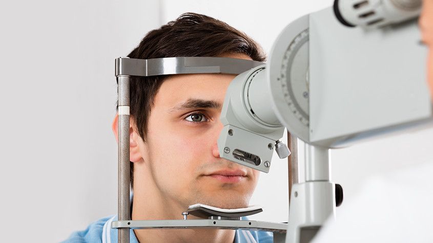
What to expect from Optomap scanning
Optomap is a fast, painless and non-invasive procedure that is suitable for patients of all ages, even children and pregnant women. Many patients require their eyes to be dilated ahead of the scan and will be given eyedrops which will widen their pupils and make it easier for the camera to see the structures inside the eye. Pupil dilation is painless, but patients may feel more sensitive to light both during their Optomap scan and afterwards for up to 24 hours. You may also have slightly blurred vision for a few hours. Once your eyes are dilated, you’ll be sat down and asked to look into a small device that will take the pictures of your retina. A short flash of light will let you know that the image has been taken, and the entire imaging is over in just a few seconds. The results will be sent digitally to your eye doctor who will then evaluate them. The results will also be stored on your personal optical record for future information.
If you would like more information about what is involved in Optomap, or to schedule an appointment for this effective screening technology, please contact our eyecare team.

Neurolens are the first and only prescription lenses that include an element of contoured prism in their design. This prism is designed to bring the patient’s eyes into more equal alignment, and this should help to provide relief from the symptoms that are associated with several eye misalignment conditions, including digital eye strain and binocular vision dysfunction.
What is digital eye strain?
Digital eye strain is the name given to describe a group of symptoms that can occur when someone spends long periods of time using digital devices. Since using digital devices requires the eyes to work harder than normal and we don’t always position our devices the perfect distance away, it can lead to issues such as eye pain, dry and irritated eyes, eye fatigue, light sensitivity and blurred vision. Unsurprisingly, the number of people who are experiencing digital eye strain has grown significantly over the last few years and is expected to continue to do so.
What is binocular vision dysfunction?
Binocular vision dysfunction, also known as BVD for short, is another eye condition but is one that is very misunderstood. Binocular vision dysfunction occurs when the eyes aren’t perfectly aligned, causing your brain and eyes to work harder than normal in order to create a clear visual image and remain focused. This places pressure on the trigeminal nerve, which is the nerve that is responsible for the majority of the sensations that we experience in our head and back. BVD can often manifest as other things owing to the huge range of symptoms that are associated with the condition. These can include, but aren’t limited to:
Blurred vision
Headaches/migraines
Double vision
Motion sickness
Vertigo
Dizziness
Anxiety
Many people don’t think to visit an eye doctor when they are experiencing these symptoms, but all can occur simply because the eyes are out of alignment.
What are Neurolens lenses and how do they help?
As well as containing your usual prescription, Neurolens lenses also contain a specific amount of contoured micro-prism. This micro-prism alters the position of images so that they are aligned in the same plane. This then reduces the pressure on the muscles around the eyes as well as bringing the eyes into alignment, easing the symptoms that the patient has been experiencing.
The amount of prism in Neurolens lenses is decided using the Neurolens eye-tracking device. This non-invasively measures the misalignment that the patient is experiencing, and this is used to form the basis for the patient’s Neurolens prescription. After this, it’s fairly normal for the amount of prism to need to be adjusted by infinitesimal amounts to achieve the optimal relief from your symptoms. Most patients who choose Neurolens treatment see a 50% improvement in their vision as soon as they start to have micro-prism incorporated into their prescription lenses. However, with careful adjustments, many patients see as much as an 80% reduction in the effects of digital eye strain and binocular vision dysfunction.
Want more information about Neurolens? Please contact our knowledgeable eye care specialists.
Dry eyes are one of the most common conditions that can affect our eyes and is estimated to affect millions of Americans. As you’ve probably guessed, dry eyes occur when tears fail to provide enough natural lubrication for the eyes to be comfortable and healthy. Exactly what causes dry eyes can vary significantly, from side effects from medications to prolonged computer use. What is clear is that while the condition isn’t sight-threatening, it can make day to day life much harder than it needs to be. Fortunately, there are treatments that can help, and arguably one of the most effective is Lipiflow.
What is Lipiflow?
Lipiflow is a new technological solution that addresses the underlying cause of your dry eyes, rather than simply treating the symptoms. It is most effective at helping patients whose dry eyes are caused by meibomian gland dysfunction – a condition characterized by problems with the way that the meibomian glands produce the oil that forms an essential part of our tear film. The meibomian glands can become less productive, or in some cases, even blocked by hardened oil deposits. This prevents the oil from reaching your tear film, making it less effective. Lipiflow targets the meibomian glands, warming them to break down oily blockages and massaging your eyes to make sure that the oil, and then the tear film, is evenly dispersed. This helps to combat the symptoms associated with dry eyes, which can include:
Eye fatigue
Dry, scratchy and uncomfortable eyes
Blurred vision
Sensitivity to light
Difficulty wearing contact lenses
Your eye doctor will be able to advise you if Lipiflow has the potential to be a suitable solution for your dry eyes.
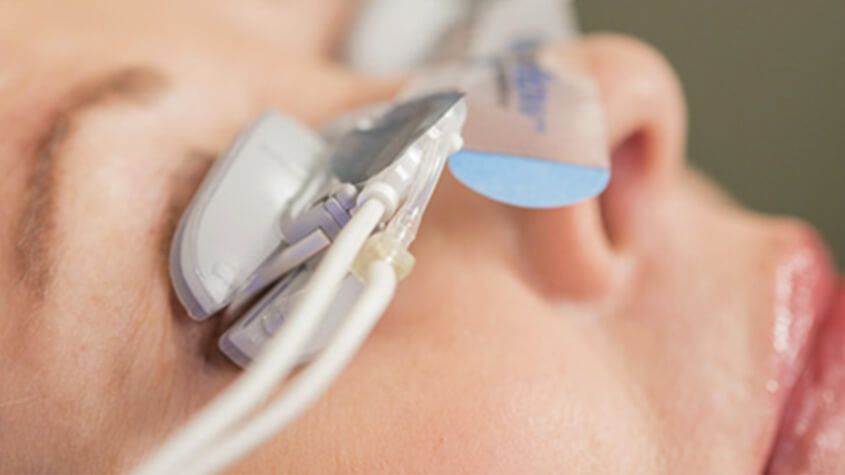
What to expect from Lipiflow treatment?
Lipiflow treatment is a simple, painless process that is performed in the comfort of your eye doctor’s office. There is no need for anesthetic. Once you are settled in your chair, your eye doctor will open the sterile, single-use applicators which are placed over your eyes. These are connected to a machine that causes the inner eyelids to heat to approximately 42.5°C to, while simultaneously placing gentle pressure on the outer eyelid surfaces. Lipiflow takes around 12 minutes per eye, during which time you can relax. You can even listen to music if you’d like to. There is no downtime, and patients can return to their usual activities right away. It takes around 3 days for patients to begin to see an improvement in their dry eye symptoms, although they may require further treatment in the future to maintain them. Optimal results are usually achieved around 6 to 8 weeks following your Lipiflow treatment.
For more information about Lipiflow, or to schedule a consultation to talk about this treatment for dry eyes, please contact our office.

If you find it difficult to tell colors apart, you may be color blind. Color blindness, or color deficiency, is estimated to affect around 8% of men and about 1% of women, but for those affected, it can significantly impact the quality of their day-to-day life. Contrary to popular belief, being color blind doesn’t mean that you can’t see any color at all. Instead, patients simply struggle to differentiate between certain colors. The vast majority of people who are color blind find it impossible to tell the difference between varying shades of red and green. You may hear this referred to as red-green color deficiency. However, this doesn’t only mean that they mix up red and green. They can also mix up colors that have some green or red light as part of their whole colors, for example purple and blue. This is because they are unable to see the red light that forms part of the color purple.
As you can probably imagine, this type of visual impairment can be a problem for things like traffic lights, taking medications and even looking at signs and directions. For example, someone who is color blind may find that the green on a traffic light may appear white or even blue.
EnChroma lens technology is specifically designed to counteract red-green color deficiency and enable patients to better identify the difference in these colors or shades. They do this by selectively filtering out the red and green wavelengths of light at the exact point where the color sensitivities overlap before hitting the retina, creating far greater contrast between the colors so that the patient can distinguish between them successfully. Most cases of color blindness respond well to EnChroma’s innovative spectral lens technology, giving patients the ability to experience life in bright, vibrant technicolor.
EnChroma lenses are made from leading edge, Trivex material, and this helps to give them the best possible quality and clarity of vision. These lenses are also extremely light, strong and offer patients 100% protection against UV light, helping to keep your eyes healthy as well as improving your vision.
If you or someone you know is color blind or color deficient and could benefit from EnChroma lenses, contact us today to learn more about how they can help!
If you are one of the thousands of people considering LASIK laser eye surgery, then you will probably be gathering as much information as possible about the treatment. By this point, you are probably aware of the benefits that LASIK offers, such as a reduced or eliminated need for glasses or contact lenses and greater convenience in your day to day life. However, for many patients, despite the advantages of LASIK, the thought of surgery on their eyes is still a cause of anxiety and fear. One of the best ways to alleviate this concern is to find out more about what the procedure entails.
Your consultation
Before you can be approved for any form of laser vision correction, including LASIK, you will need to attend a consultation appointment with your surgeon. During the consultation, he will perform an examination of your eyes and use your medical and ocular history to determine if you are a good candidate for the procedure. He will also speak to you about the expected outcome from your surgery, making you aware that while LASIK will dramatically improve your eyesight, there is no guarantee that you will not need to wear glasses in some situations, such as while driving in the dark.
How LASIK Works
LASIK uses a cool, ultraviolet beam of light to reshape the patient’s cornea. Doing so will more accurately focus the light that enters the eye on to the retina, thus improving the patient’s vision. The way in which the cornea needs to be reshaped will depend on the visual needs of the patient. For example, a patient who is far-sighted will need their cornea reshaping to be steeper to experience better eyesight. Alternatively, a patient who is near-sighted will require their cornea to be flattened in order to improve their vision. LASIK can also smooth an irregular cornea into a more standard shape, meaning that the procedure can also be used to correct astigmatism.
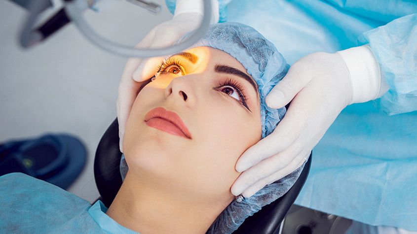
The LASIK procedure
The LASIK procedure is very fast and straightforward. Although you will probably be in the surgical suite for around half an hour, the actual process only takes a couple of minutes per eye. The rest of the time will be spent preparing and ensuring that you are comfortable. Anesthetic eye drops are given to patients before their procedure so that the entire process is pain-free. If you are particularly anxious, it may also be possible for you to be slightly sedated – this should be discussed with your doctor at your consultation appointment.
Once you are in position, we will use a femtosecond laser to cut a thin, circular flap into the outer cornea. This can then be pulled back to reveal the underlying corneal tissue, known as the stroma so that it can be reshaped using the laser. The exact path that the laser needs to take, known as the topography, will have been pre-programmed ahead of the procedure and can be followed with complete precision and accuracy.
Once the reshaping is complete, the flap is replaced back over the eye and the surgery is complete. There is no need for sutures or bandages as the cornea will start to heal immediately and without any medical intervention.

If you already rely on wearing glasses or contact lenses to be able to see clearly, you may be frustrated with the effect that they have on your life. Regular vision tests, finding glasses to suit your face shape, having to remember to take eyeglasses with you wherever you go, prescription sunglasses, fiddly contact lenses… the list of inconveniences associated with conventional ocular solutions is extensive.
LASIK is a modern, minimally-invasive procedure that can substantially reduce or eliminate your need to use eyeglasses or contact lenses, allowing you to enjoy life without limitations or inconvenience. The popularity and success of LASIK laser eye surgery have helped to make it the number one elective surgery across the globe.
Candidacy for LASIK
LASIK has an extremely high success rate. According to the American Society of Cataract and Refractive Surgery, 96% of patients achieve 20/20 vision or better. However, it’s high success rate doesn’t make LASIK automatically the right solution for everyone.
Candidacy for LASIK is assessed by through our office on a case by case basis so that you can be certain that whatever treatment is recommended for you, it will give you the very best opportunity to improve your vision. During your consultation, we will perform a thorough examination of your eyes and vision, ask you about your general health and talk you through both the procedure and aftercare.
The general guidelines for LASIK candidacy state that patients must:
be at least 18 years of age
have had stable vision with no prescription changes for a minimum of 12 months
have a current prescription for eyeglasses or contact lenses that falls between specified parameters recommended for LASIK
have no significant medical or eye-related problems such as glaucoma, or diabetic retinopathy or certain autoimmune diseases
have no history of corneal disease
not be pregnant or nursing at the time of the procedure or planning pregnancy within 6 mo.
A refraction is usually performed as a part of a routine eye examination. The purpose of this test is to determine if a person has a refractive error which would then mean the patient would need glasses or contact lenses.
What Is The Normal Value for a Refraction?
A value of 20/20 is normal (optimum) vision. This means that individuals who have 20/20 vision are able to read letters that are 3/8-inch (1 centimeter) tall from 20 feet (6 meters) away. The normal uncorrected vision (without glasses or contact lenses) refractive error is zero (plano). Individuals who don’t have 20/20 vision, have a refractive error. This means that the light passing through the eye does not focus on the retina (back of the eye) because it is either bending to little or too much. The refraction tells us what prescription lens is needed in to move the focus onto the retina to provide 20/20 vision.
For people over age 40 who have normal distance vision but difficulty with near vision, a refraction with a small type size is used to determine normal near vision and the correct power for reading glasses.
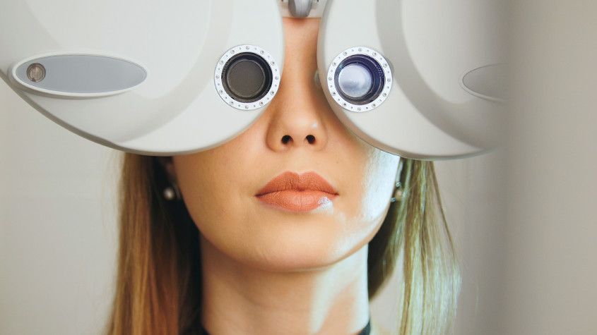
How Is A Refraction Performed?
The test is performed by having the patient look through a special device called a phoropter. The patient looks through the phoropter and focuses on an eye chart 20 feet (6 meters) away. The phoropter contains hundreds of lenses of different strengths that are shown to the patient to provide the clearest vision. The test is performed one eye at a time. If the patient is wearing contact lenses, they should be removed before the test.
In case the final vision is less than 20/20 even with lenses, then this is a indication that there may be some underlying pathology, which would warrant further testing.
What Can Cause a Refraction other than Plano?
Astigmatism (abnormally curved cornea causing blurred vision)
Hyperopia (farsightedness)
Myopia (nearsightedness)
Presbyopia (inability to focus on near objects that develop with age)
All conditions above should be able to obtain 20/20 vision with glasses or contacts
Conditions below that may not be able to obtain 20/20 vision with a refraction
Corneal ulcers and infections
Loss of sharp vision due to macular degeneration
Retinal detachment (separation of the light-sensitive membrane (retina) in the back of the eye from its supporting layers)
Retinal vessel occlusion (blockage in a small artery that carries blood to the retina)
Retinitis pigmentosa (an inherited disorder of the retina)
There is an art to refraction and we will always answer the your questions and as well as discuss their findings. Based on the results of the refraction, we can determine the amount of myopia, hyperopia or astigmatism.
The parts of a comprehensive eye examination vary according to the patient's age, date of last exam, and other factors. Not all parts of the eye exam may be needed or performed, but the first part of the eye exam will include documenting medical history. Here are some eye and vision tests that are likely to be encountered during a comprehensive eye exam:
Visual Acuity Tests
Visual acuity tests measure the sharpness of vision and are usually performed using a projected eye chart to measure the distance visual acuity and a hand-held small acuity chart to measure the near vision (for reading).
Color Blindness Test
A screening test that checks the color vision is often performed early in a comprehensive eye exam to rule out color blindness.
Cover test to check eye alignment.
A test used to assess strabismus or a more subtle binocular vision problem that could cause eye strain or amblyopia (lazy eye).
Ocular Motility (Eye Movements) Testing
Ocular motility testing is performed to determine how well eyes can follow a moving object and/or quickly move between and accurately fixate on two separate targets.
Stereopsis (Depth Perception) Test
This is used to test perception of depth and 3-dimensional structure obtained on the basis of visual information deriving from two eyes by individuals with normally developed binocular vision.
Retinoscopy
This test is used to estimate which lens powers will best correct distance vision. Based on the way the light reflects from the eye, the doctor is able to obtain an approximation of the eyeglass prescription. This test is useful for children and patients who are unable to accurately answer the doctor's questions.
Manual refraction with a phoropter.
This is the test used to determine the exact eyeglass prescription.

Macular degeneration, commonly referred to as age-related macular degeneration (AMD), is the single largest cause of sight loss in the developed world and affects more than 10 million Americans. It usually affects people over the age of 60, but has been known to affect those who are younger. It is a painless condition that usually affects both eyes with the loss being experienced in the central vision. It does not affect the peripheral vision, meaning that it does not cause total blindness.
What is the macula?
The macula is the most sensitive part of the retina and is responsible for our central vision and what allows us to see fine details with clarity.
Varieties of AMD
Wet AMD
Wet AMD is one variety of the condition in which abnormal blood vessels grow into the macula, leaking blood or fluid which then causes scarring and a rapid loss of central vision. Wet AMD can develop suddenly and rapid referral to a specialist is essential as it can be treated if caught quickly.
Dry AMD
Dry AMD is the most common variety of age-related macular degeneration and is a gradual deterioration of the retina as the cells die off over time and are not regenerated. Up to 15% of people with dry AMD go on to develop wet AMD, and so any sudden changes in your vision should be followed up with your optometrist as soon as possible.

When you were a kid, did you experience your eyes become reddish and all of a sudden, someone close to you was also suffering from it? Your eyes, as well as those who contracted it, got itchy and swollen, right? Then it must have been that you were suffering from pink eye.
Pink eye is well known as conjunctivitis and it is the infection or inflammation of the conjunctiva or the transparent membrane that serves as a covering for the white part of the eye called the sclera that lines the eyelid. In addition to inflammation, there is usually tearing in the eyes that emits a sticky discharge which develops into a crust while one is sleeping, making it difficult for the patient to open their eyes in the morning.
One thing about the pink eye, which could affect one or both eyes, is that it is highly contagious. While it is more common in children, adults can also victims of this eye condition. Here are the 3 major causes of pink eye:
Bacteria
Streptococci and staphylococci are bacteria types that are most responsible for pink eye. However, chlamydia and gonococci can also cause pink eye. It is accompanied by serious eye pain, itching, swelling, redness, and discharge. The spread of bacterial pink eye is usually as a result of using personal items of infected parties, such as makeup or makeup tools that have been infected with bacteria or putting dirty hands in the eyes. If not treated, it can last for more than 10 days, but if treated, it should resolve in less than 3 days.
Allergies
Pink eye caused by allergies is followed by serious itching and tearing of the eyes. Pain is minimal, but it typically comes with quite a bit of discomfort. Most of the time, pink eye is accompanied by sneezing or coughing. Allergens that trigger pink eyes include grass, dust, pollen, mold, and ragweed. Allergy based pink eye is not usually contagious.
Viruses
Viruses such as the adenoviruses and herpes virus are the most common causes of pink eye. When a virus is the cause, there is usually a lot of teary discharge accompanied by nasal congestion, puffy eyelids, runny nose, and sharp pain. It is usually contracted from cough and sneeze droplets from an infected individual. It can take as long as 2 weeks to treat depending on the seriousness of the infection.
General Symptoms of Pink Eye:
Itchy eyes
Redness of the sclera
Pain
Watery discharge
Swollen eyelids
Hazy or blurry vision
Oversensitivity to light
Prevention
The best way to prevent pink eye is by practicing good hygiene which includes:
Avoid putting dirty hands in your eyes
Make it a habit to wash your hands often
Avoid sharing towels and other personal items
Do not use dirty items
Changing your pillow cover regularly
Do not leave a makeup item open for too long
Avoid sharing makeup items like eyeliners, mascara, etc.
Treatment of Pink Eye
The treatment of pink eye is dependant on its underlying cause. If it is caused by a virus, you just might have to wait for the virus to run its course which could last for about four to seven days. Virus caused pink eye could be easily contracted so it is imperative to try and prevent further spreading. Viruses cannot be cured by antibiotics, but some antiviral drugs could be helpful.
Antibiotics are most effective against pink eye caused by bacteria as they reduce the lifespan of these bacteria and could come in the form of eye drops or pills. Based on the doctor’s prescription, an eye drop should be administered about four to six times daily. It is important you finish using your drugs even after the disappearance of symptoms.
To deal with pink eyes caused by allergies, the allergy should be treated. Once treated, pink eye should disappear. It is also important to avoid allergens as much as possible so as to avoid pink eye.
Whenever the symptoms of pink eye emerge, the best preventive measure is to stay at home until the watery discharge ceases to avoid the spread of the bacteria or virus. You should also visit your doctor immediately to begin treatment. While mild pink eyes generally go away on its own, some of the more serious forms can cause a scar on the cornea.


© 2024 Skowron Eye Care. All rights Reserved. Accessibility Statement - Privacy Policy - Sitemap
Powered by: